
Why the FNA vs Biopsy Question Comes Up
When your veterinarian feels a lump or finds a mass on imaging, the next step is figuring out what it is. Two procedures dominate that decision, and the fna vs biopsy pets question is one of the most common crossroads in veterinary oncology. A fine-needle aspirate (FNA) collects loose cells through a thin needle, while a biopsy removes a piece of tissue with its architecture intact. Both send samples to a pathologist, but they answer questions at different depths.
In this article
- Why the FNA vs Biopsy Question Comes Up
- What a Fine-Needle Aspirate Shows
- What a Biopsy Shows
- The Honest Tradeoffs in the FNA vs Biopsy Pets Decision
- When Each Test Is Chosen
- What the Sample Report Means for You
- Cost and Access Considerations
- Diagnosis as a Starting Point, Not a Verdict
- Frequently Asked Questions
Choosing between them is not about which test is “better” in the abstract. It is about which test answers the question your team needs answered, with the least cost and risk that still gets a reliable result. Sometimes an FNA is plenty. Sometimes only a biopsy will do. Often the two are used in sequence.
Understanding the tradeoffs helps you have a calmer, clearer conversation with your veterinarian or a board-certified oncologist (DACVIM-Oncology) and the pathologist (DACVP) who reads the slides.
What a Fine-Needle Aspirate Shows
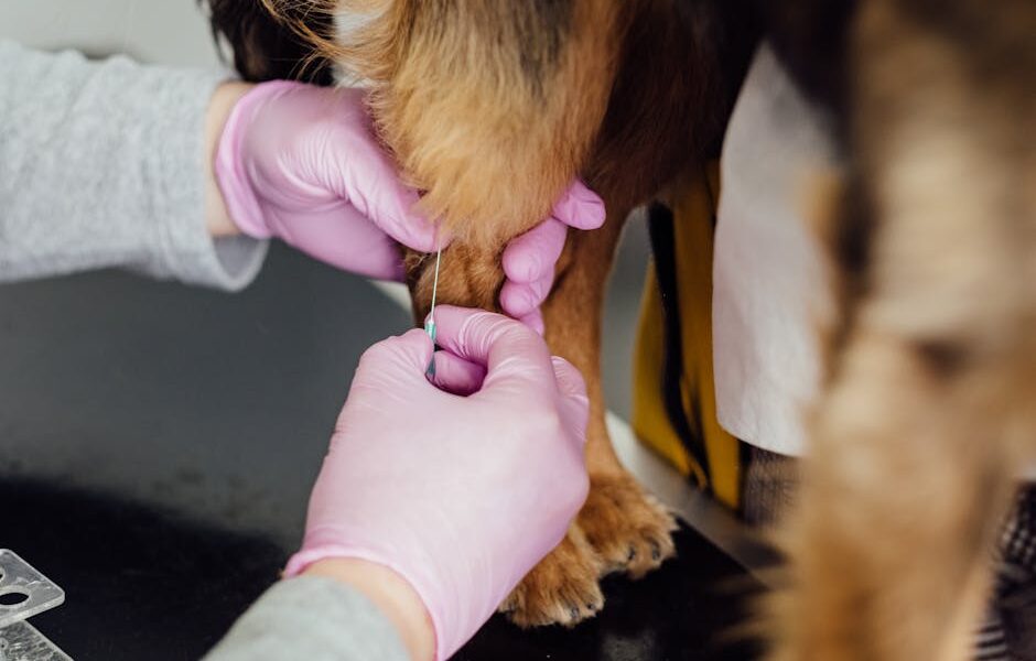
An FNA uses a small needle, often the size used for a vaccine, to pull a sample of individual cells from a mass. Those cells are smeared on a slide, stained, and examined under a microscope in a process called cytology. The procedure is quick, usually needs no sedation, and rarely requires more than a moment of restraint.
Cytology can be remarkably informative. For many tumors, such as lipomas, many mast cell tumors, and several round-cell cancers, an FNA gives a confident answer. It can also screen lymph nodes and organs for spread during staging. Because it is low-cost and low-risk, the FNA is often the very first diagnostic step.
Its limit is that it shows cells without their surrounding structure. A pathologist sees what the cells look like but not how they are arranged, which matters for some diagnoses. An FNA can also return an inconclusive result if too few cells are collected or the tumor does not shed cells easily.
Some tumors aspirate beautifully and others barely at all. Round-cell tumors, such as mast cell tumors, lymphoma, and histiocytomas, tend to shed cells readily and are often well suited to cytology. Many sarcomas and some hard, fibrous masses give up few cells, so an FNA may come back unrewarding even when the technique was flawless. Knowing this in advance helps set expectations: an inconclusive FNA on a firm sarcoma is common and is not a sign that anything went wrong.
What a Biopsy Shows
A biopsy removes a piece of tissue rather than scattered cells, preserving the architecture so the pathologist can see how cells relate to one another, to blood vessels, and to surrounding tissue. This is called histopathology, and it is the gold standard for a definitive diagnosis. Our guide to reading the histopathology report walks through the grade and margin details a biopsy can reveal.
Biopsies come in several forms. An incisional biopsy takes a wedge of a larger mass. An excisional biopsy removes the whole lump. A needle-core biopsy uses a wider hollow needle to grab a small cylinder of tissue. Each balances how much tissue is collected against how invasive the procedure is.
The tradeoff is that biopsies are more involved. They usually require sedation or anesthesia, can bleed or become infected, and cost more. But for tumors where grade and architecture drive treatment, that extra information is worth it.
There is also a strategic question of where to biopsy. For a mass that will likely need surgery, the surgeon often plans the biopsy tract so it can be removed entirely with the tumor later, avoiding seeding healthy tissue. This is one reason a biopsy is best coordinated with whoever may operate, rather than done in isolation. A poorly placed incisional biopsy can complicate a curative-intent surgery down the line.
Excisional Biopsy: Diagnosis and Treatment at Once
For small, accessible masses where removal is low-risk regardless of the diagnosis, an excisional biopsy can be efficient: the whole lump comes off and is sent for analysis in a single step. The catch is that if it turns out to be an aggressive tumor needing wide margins, the first surgery may not have removed enough, and a second, larger operation could follow. For that reason, excisional biopsy is favored when the suspected diagnosis is benign or low-risk, and a diagnosis-first approach is favored when an aggressive tumor is on the table.
The Honest Tradeoffs in the FNA vs Biopsy Pets Decision
The core of the fna vs biopsy pets decision is a set of genuine compromises, and a good team will name them plainly. FNA wins on speed, cost, and minimal risk, but it can be inconclusive and cannot always grade a tumor. Biopsy wins on diagnostic certainty and grading but costs more, needs anesthesia, and carries small surgical risks.
- Certainty: Biopsy gives a more definitive diagnosis; FNA may be suggestive rather than conclusive.
- Invasiveness: FNA is nearly painless and awake; biopsy usually needs sedation or anesthesia.
- Cost: FNA is inexpensive; biopsy adds anesthesia, surgical, and lab fees.
- Information depth: Only a biopsy reliably reports grade, margins, and tissue architecture.
There is no universally right answer. A small skin lump in a healthy young dog might start with an FNA, while a suspected aggressive sarcoma in a planning-for-surgery patient may go straight to a biopsy.
When Each Test Is Chosen
Veterinarians generally reach for an FNA first when a mass is easy to access, when a quick answer would guide the plan, or when screening lymph nodes and organs during staging. It is the sensible opening move for most superficial lumps and many internal masses sampled under ultrasound guidance.
A biopsy is chosen when the FNA was inconclusive, when the tumor type cannot be confirmed on cytology, or when the team needs the grade to plan surgery, chemotherapy, or radiation. For tumors like soft tissue sarcomas, our overview of sarcoma grading and surgical margins shows why architecture-level detail changes the surgical approach. The sequence is logical: start least invasive, escalate only as the questions require.
Image-Guided Sampling
For masses inside the chest or abdomen, both aspirates and core biopsies are frequently collected with ultrasound or CT guidance. This lets the team sample a deep organ precisely while watching the needle in real time, improving accuracy and safety.
What the Sample Report Means for You
A cytology or biopsy report can feel intimidating, so it helps to know the common outcomes. A cytology report may say “consistent with” a tumor type, which signals a strong but not absolute conclusion, or “non-diagnostic,” meaning too few usable cells were collected and another sample is needed. A histopathology report will typically name the tumor, often with a grade and, after surgery, a comment on margins.
The most important habit is to read the report alongside your veterinarian rather than alone. Ask whether the result is definitive or suggestive, whether further sampling is recommended, and how the finding changes the plan. The pathologist interprets the slides, but your team translates that into decisions for your individual pet.
It is worth understanding that a cytology and a biopsy can sometimes disagree, and when they do, the biopsy generally carries more weight because it preserves architecture. If your pet’s FNA suggested one thing and a later biopsy says another, that is not a contradiction so much as a more detailed test refining a preliminary one. The same is true when an FNA is reassuring but the mass keeps growing: behavior that does not match the cytology is a reason to escalate to a biopsy rather than to assume the first result was final.
Cost and Access Considerations
An FNA with in-house or send-out cytology is among the most affordable diagnostics in veterinary medicine, often a modest add-on to an exam. A surgical biopsy, with anesthesia, the procedure itself, and histopathology, costs considerably more and varies widely by region and facility, current as of 2024 to 2025. Specialty interpretation and immunohistochemistry can add further expense.
If finances are tight, ask your team to prioritize the test that will actually change the plan, and look into financing options such as CareCredit or Scratchpay. Skipping diagnostics to save money sometimes costs more later when treatment proceeds without a clear target.
Diagnosis as a Starting Point, Not a Verdict
A cancer diagnosis confirmed by FNA or biopsy can feel like the end of the road, but for many pets it is the beginning of a plan. Many families adopt or keep pets with cancer histories and give them wonderful lives. If you are weighing a commitment, our guide to adopting a pet with a chronic illness offers a grounded, hopeful perspective.
The point of choosing the right sampling test is to replace fear with information. Whether the answer comes from a needle or a scalpel, knowing what you are facing is the first step toward caring for your pet well.
Frequently Asked Questions
Is a fine-needle aspirate painful for my pet?
Most pets tolerate an FNA with little more than the brief discomfort of a vaccine. It uses a thin needle, takes seconds, and rarely needs sedation. Deeper, image-guided aspirates may use light sedation for comfort and accuracy.
If the FNA already gave an answer, why does my vet still want a biopsy?
Cytology can identify many tumors but cannot always grade them or confirm certain types. If the grade will guide surgery, chemotherapy, or radiation, a biopsy provides the tissue-level detail an FNA cannot, so the recommendation reflects what the plan needs.
Can a biopsy spread the cancer?
For most tumors this risk is very low and is far outweighed by the value of an accurate diagnosis. For a few specific tumor types, surgeons use careful technique to minimize any theoretical seeding. Discuss any concerns with your oncologist or surgeon.
What does a non-diagnostic result mean?
It means the sample did not contain enough usable cells or tissue to reach a conclusion, not that something was done wrong. The next step is usually to repeat the FNA, use image guidance, or move to a biopsy for a larger sample.
How long do results take?
Cytology from an FNA can sometimes be read same-day in-house or within a few days at a reference lab. Histopathology from a biopsy typically takes several days to over a week, and added tests like immunohistochemistry extend that timeline. Your veterinarian can give a facility-specific estimate.


