
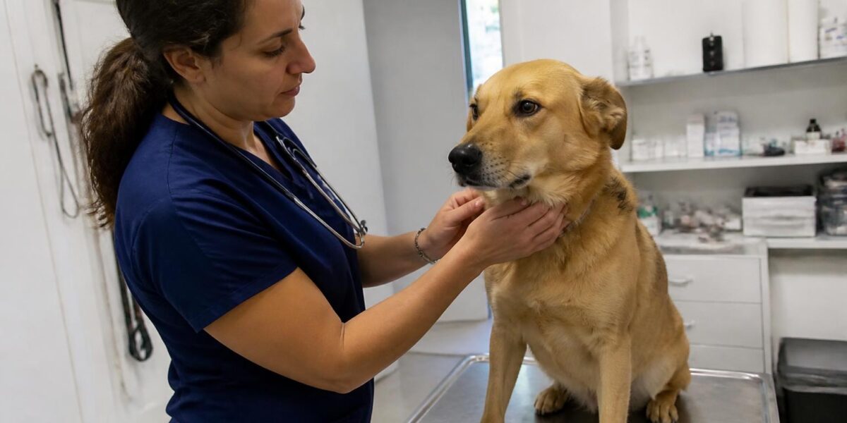
What Idiopathic Facial Nerve Paralysis in Dogs Is
Idiopathic facial nerve paralysis in dogs is a sudden loss of function of the facial nerve (cranial nerve VII) that occurs without an identifiable cause. The facial nerve controls the muscles that close the eye, move the lips and ear, and produce some tear and saliva flow. When it stops working, owners notice a one-sided facial droop that can look alarming but is, in many cases, less serious than it appears.
In this article
- What Idiopathic Facial Nerve Paralysis in Dogs Is
- Recognizing the Clinical Signs
- Differentiating From Other Facial Conditions
- The Diagnostic Workup
- Treatment of the Underlying Cause
- Idiopathic Cases and Conservative Management
- Eye Care: The Most Important Home Task
- Prognosis and Long-Term Outlook
- Frequently Asked Questions
The condition is most commonly seen in middle-aged to older dogs, with a documented predisposition in Cocker Spaniels and a few other breeds. Onset is typically sudden — owners often notice the change overnight. The diagnosis is one of exclusion, made after ruling out treatable causes like otitis media, hypothyroidism, neoplasia, and trauma.
Many affected dogs improve gradually over weeks to months, though some are left with residual facial asymmetry. The most important short-term concern is corneal protection — without normal blinking, the eye on the affected side can dry out and ulcerate.
Recognizing the Clinical Signs
Owners typically notice a combination of facial signs. The lip on the affected side droops and may drool food and water. The ear hangs lower than the other. The eye may not blink normally — owners notice the eye stays open, looks dry, or develops discharge. The nose may pull slightly toward the unaffected side.
The eye changes are the most clinically important to recognize quickly. Without normal blinking and tear film distribution, the cornea dries out and can develop ulcers within days. Persistent squinting, redness, cloudiness, or discharge in the affected eye warrants prompt veterinary attention regardless of the broader workup timeline.
Because vestibular signs sometimes accompany facial nerve paralysis when the underlying cause is in the inner or middle ear, watch also for head tilt, ataxia, or rapid eye movements (nystagmus). The combination of facial paralysis plus vestibular signs strongly suggests an otic cause and warrants thorough imaging.
Differentiating From Other Facial Conditions
Facial droop has several possible causes, and the diagnostic workup is designed to distinguish among them. Otitis media in dogs — middle ear infection — is one of the most important treatable causes and can produce facial nerve paralysis through inflammation around the nerve as it passes through the middle ear cavity. Treating the otitis often allows facial nerve function to recover.
Hypothyroidism in dogs is associated with peripheral neuropathies including facial nerve paralysis in some patients. Bloodwork that includes a full thyroid panel is part of the standard workup for any new facial nerve paralysis. Treating the hypothyroidism may improve the neurologic signs over weeks to months.
Other differentials include neoplasia of the middle ear or skull base, trauma, inflammatory polyps, tickborne infections in endemic areas, and rare conditions like masticatory muscle myositis (which involves chewing muscles rather than facial nerve function). Idiopathic facial nerve paralysis is the diagnosis of exclusion when imaging and bloodwork find nothing.
The Diagnostic Workup
Your primary veterinarian will start with a thorough history and a complete neurologic exam, paying particular attention to cranial nerve function on both sides of the face. A careful otoscopic examination evaluates the visible ear canal and tympanic membrane. Bloodwork including a CBC, chemistry panel, and full thyroid panel screens for systemic disease.
If the otoscopic exam suggests middle ear involvement or if signs are progressive, advanced imaging is the next step. CT or MRI of the head, performed by a board-certified ACVIM-Neurology specialist or ACVR radiologist, evaluates the middle ear cavities, the path of the facial nerve, and the surrounding structures for masses, fluid, or other abnormalities.
In endemic areas, tickborne disease titers may be appropriate. If the eye is affected, your vet will also assess tear production with a Schirmer tear test and examine the cornea with fluorescein staining to detect any early ulceration. Comprehensive workup costs add up quickly, so review the veterinary specialist cost breakdown alongside your pet insurance decision for dogs guide.
Treatment of the Underlying Cause
When a specific cause is identified, treatment targets it directly. Otitis media is treated with appropriate long-course antibiotics, sometimes a myringotomy to drain the middle ear, and management of any underlying ear canal disease. Hypothyroidism is treated with thyroid hormone replacement and monitored with periodic bloodwork. Masses identified on imaging are evaluated for biopsy and treated according to the histopathology.
For tickborne infections in endemic areas, appropriate antibiotic therapy is started after testing. For trauma, supportive care plus management of any associated injuries is the approach. The principle is the same in every case: identify what is interrupting the facial nerve, treat it, and monitor the nerve function for gradual recovery.
Recovery of facial nerve function takes time even after the underlying cause is treated — weeks to months is typical. Some patients are left with residual asymmetry; others recover completely. Patience and follow-up examinations are essential during this period.
Idiopathic Cases and Conservative Management
When advanced imaging and bloodwork are unrevealing, the diagnosis defaults to idiopathic facial nerve paralysis. There is no specific treatment for idiopathic cases — management focuses on protecting the affected eye, supporting general comfort, and monitoring for spontaneous improvement over weeks to months.
Many idiopathic cases improve at least partially over weeks to months, though some retain residual asymmetry. A subset of dogs experience the condition on the opposite side weeks or months later, which is unusual but documented. Each new episode warrants a fresh recheck because not every recurrence stays idiopathic — sometimes a previously hidden cause becomes apparent.
Owners often want to “do something” but the right intervention is patient supportive care plus frequent monitoring of the eye. Resist the urge to seek experimental therapies; idiopathic cases follow their own course regardless of unproven treatments.
Eye Care: The Most Important Home Task
The single most important home task during recovery is corneal protection. Without normal blinking, the affected eye dries out and is at high risk for ulceration. Your vet will prescribe artificial tear ointment or solution to be applied multiple times daily — frequency varies, but every two to four hours during waking hours is typical for severely affected eyes.
Watch closely for any signs of corneal trouble: squinting, increased discharge, cloudiness, redness, or a visible defect on the surface. Any of these warrants same-day or next-day veterinary evaluation. A corneal ulcer in a poorly blinking eye can progress quickly.
Some dogs benefit from a soft cone or face shield to prevent self-trauma during the recovery period. Others tolerate the eye drops well and need no further protection. Tailor the home setup to your individual dog and check in with your vet at the first sign of worsening.
Prognosis and Long-Term Outlook
Prognosis for idiopathic facial nerve paralysis is generally favorable, though recovery is gradual and incomplete in some patients. Most dogs adapt well to residual facial asymmetry and continue to enjoy a full quality of life. The main long-term concern is the eye on the affected side, which may require ongoing artificial tear support if blinking does not fully recover.
For dogs with a treatable underlying cause, prognosis depends on the cause itself. Otitis media-related cases often recover well with appropriate antibiotic therapy. Hypothyroid-related cases may improve over weeks to months on thyroid hormone replacement. Tumor-associated cases vary widely depending on tumor type, location, and treatment options.
Coordinated care between your primary vet and a board-certified ACVIM-Neurology specialist gives the best long-term outcomes. Recheck examinations at four-week intervals during active recovery help catch any complications and track progress. Many dogs return to essentially normal lives, with only a slightly different smile to remind owners of their journey.
Frequently Asked Questions
Is idiopathic facial nerve paralysis painful?
The paralysis itself is not painful. Pain, when present, often points to an underlying cause like otitis media. Your vet will assess for and treat any associated pain during the diagnostic workup and follow-up appointments.
How long does recovery take?
Recovery timelines vary widely. Some dogs improve within weeks; others take months and may have residual asymmetry. When a treatable underlying cause is found and addressed, improvement often follows over weeks to months. Patience and consistent eye care are key.
Can my dog still eat and drink normally?
Most dogs adapt quickly to facial weakness for eating and drinking. Some drool more than usual on the affected side; some food may fall out of the mouth temporarily. Soft food and slightly elevated bowls help. If your dog is not eating or losing weight, contact your vet promptly — that is a different concern.
Will my dog need an MRI?
If otoscopic exam, bloodwork, and clinical assessment do not reveal an obvious cause, advanced imaging is usually recommended to rule out serious treatable conditions. CT and MRI both have roles depending on what your vet is looking for. Your ACVIM-Neurology specialist will choose the right modality for your dog.
Can it happen on the other side later?
Yes, though it is unusual. A small subset of dogs experience facial nerve paralysis on the opposite side weeks or months later. Each new episode warrants a fresh diagnostic workup because not every recurrence is idiopathic.


