
What Mast Cell Tumors Are
Mast cell tumors are among the most common skin cancers in dogs, accounting for a significant share of canine skin tumors overall. They originate in mast cells, a type of immune cell that normally helps with allergic responses and parasite defense. When mast cells become cancerous, they form lumps that can range from harmless and easily cured to aggressive and life-threatening.
In this article
Mast cell tumors dogs develop are notorious for their variable appearance. They can look like virtually any other skin lump: small bumps, soft fatty-feeling masses, raised pink lesions, or even ulcerated wounds that will not heal. Because of this variability, all skin lumps in dogs deserve veterinary evaluation rather than assumption that they are benign.
Several breeds are at higher risk, including Boxers, Boston Terriers, Bulldogs, Pugs, Labrador Retrievers, Golden Retrievers, and Pit Bull-type dogs. Mixed-breed dogs can certainly develop mast cell tumors too. The disease is more common in middle-aged to older dogs, although it can appear at any age.
The Importance of Aspirate or Biopsy
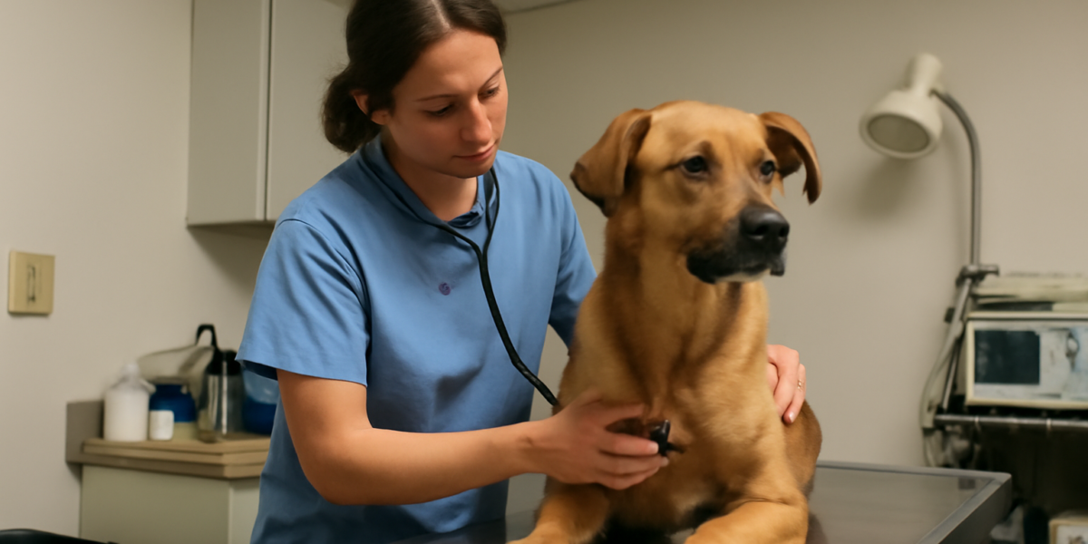
Diagnosis starts with fine needle aspiration of the suspicious lump. The veterinarian inserts a small needle into the mass and collects cells, which are examined under a microscope. Mast cells have a characteristic appearance, with dark granules visible on stained slides, that usually allows quick diagnosis.
Once a mast cell tumor is identified, additional testing helps assess behavior. Surgical biopsy provides definitive grading, which guides treatment decisions and prognosis. Some tumors are graded after removal because pre-surgical grading from aspirate alone is not always reliable.
Staging tests assess whether the tumor has spread. These typically include lymph node aspiration, bloodwork, abdominal ultrasound, and sometimes chest X-rays or other imaging. Early-stage low-grade tumors may need only minimal staging, while higher-grade tumors warrant more thorough evaluation. Your veterinarian will tailor the workup to your dog.
The Grading System
Mast cell tumors are traditionally graded I, II, or III on the Patnaik system, with grade I being least aggressive and grade III most aggressive. A newer two-tier system (low-grade vs high-grade Kiupel system) is increasingly used because it correlates better with behavior and is more reproducible between pathologists.
Low-grade tumors typically behave well. They tend to be slow-growing, less likely to spread, and often cured by complete surgical removal. Many low-grade tumors never cause additional problems once removed with adequate margins. The prognosis for these dogs is generally excellent.
High-grade tumors are more aggressive. They are more likely to spread to lymph nodes, abdominal organs, or other sites, and often require additional treatment beyond surgery. Even with comprehensive treatment, prognosis is more guarded. Your veterinarian or a board-certified oncologist will discuss expectations based on the specific grade and stage.
Surgery as First-Line Treatment
Surgery is the cornerstone of mast cell tumor treatment. Wide local excision, where a generous margin of normal-appearing tissue is removed around the tumor, gives the best chance of complete removal. Margins are typically two to three centimeters in all directions plus one tissue plane deep, although the exact approach depends on tumor location and grade.
Some tumors in challenging locations (like the limbs or face) cannot be removed with such wide margins without significant tissue loss. In those cases, the surgeon does the best they can and additional treatments fill in the gaps. Reconstructive techniques can help close challenging surgical sites.
Pathology evaluation of the removed tissue is essential. The pathologist confirms the grade, assesses whether margins are clean (complete excision) or dirty (cancer cells remain), and provides additional information that guides next steps. Clean margins on a low-grade tumor often mean treatment is complete.
Adjuvant Treatments
When surgical margins are dirty or the tumor is high-grade, additional treatments are often recommended. Radiation therapy targets the surgical site to kill any remaining cancer cells. Radiation requires referral to a specialty hospital and multiple treatment sessions, but outcomes are good for localized disease.
Chemotherapy is used for high-grade tumors, especially those that have spread to lymph nodes or other sites. Common drugs include vinblastine, lomustine, and prednisone, often used in combination protocols. Newer targeted therapies like toceranib (Palladia) and masitinib block specific cell signals and have transformed treatment for some dogs.
The right treatment combination depends on grade, stage, location, your dog’s overall health, and family logistics. Your veterinary oncologist will help develop a tailored plan. Many dogs do extremely well with appropriate adjuvant therapy, even with high-grade disease.
Mast Cell Granule Release
One unique feature of mast cell tumors is that they can release the inflammatory granules stored in mast cells. These granules contain histamine, heparin, and other compounds that cause local inflammation, GI ulceration, and sometimes systemic symptoms. The release can happen spontaneously, with handling, or during surgery.
Local effects include swelling, redness, and itching at and around the tumor. Tumors can suddenly become inflamed and ulcerated, which is sometimes called Darier’s sign. Pre-treatment with antihistamines (H1 blockers like diphenhydramine and H2 blockers like famotidine) is often used before surgery to prevent dramatic reactions.
Systemic effects include GI ulceration with vomiting, decreased appetite, and dark tarry stools (melena). These signs warrant prompt veterinary attention. Long-term GI protection with H2 blockers or proton pump inhibitors is sometimes recommended for dogs with high-grade disease or systemic involvement.
The Targeted Therapy Era
The development of targeted therapies has changed mast cell tumor treatment significantly. Toceranib (Palladia) and masitinib (Kinavet, less commonly available) target specific cell signals, including the c-KIT mutations found in some mast cell tumors. Dogs with c-KIT mutations often respond particularly well to these drugs.
Toceranib is given orally several times per week and is generally well-tolerated. Side effects include GI upset, decreased appetite, and occasional bloodwork changes. Periodic monitoring during treatment helps catch issues early. Many dogs respond meaningfully, with tumors shrinking or stabilizing for extended periods.
Genetic testing for c-KIT mutations on tumor samples can help predict response to targeted therapy. Mutation-positive tumors often respond best, although tumors without the mutation can still respond. Your veterinarian or oncologist will discuss whether testing is worthwhile for your dog’s situation.
Long-Term Monitoring
Dogs who have had mast cell tumors are at increased risk for developing additional ones, sometimes years later. Regular skin checks at home and during veterinary visits catch new lumps early. Any new mass deserves prompt evaluation, ideally with fine needle aspiration to confirm or rule out mast cell origin.
Recheck appointments after initial treatment include physical exams, palpation of regional lymph nodes, and sometimes imaging or bloodwork. The frequency depends on tumor grade and stage. Higher-grade tumors require more intensive monitoring, while low-grade tumors with clean margins may need less frequent follow-up.
For more on canine cancer topics, our guide to lymphoma in dogs covers another common cancer. Comprehensive senior wellness exams and prompt evaluation of any new lumps are essential parts of long-term care for dogs with cancer history.
Quality of Life Considerations
Most dogs treated for mast cell tumors enjoy good quality of life. Low-grade tumors that are completely removed often have no further impact. High-grade tumors with appropriate adjuvant treatment can result in meaningful additional time with good quality of life.
Watch for signs that warrant veterinary attention: new lumps, sudden changes in existing masses (size, color, ulceration), GI symptoms (vomiting, decreased appetite, dark stools), lethargy, or any concerning systemic signs. Mast cell disease can flare unexpectedly, and prompt response prevents complications.
For broader canine wellness topics and adoption resources, our dog care blog and dog adoption hub have helpful information. Many dogs with cancer history make wonderful companions and only need attentive monitoring.
Prevention and Early Detection
Mast cell tumors cannot be reliably prevented, but early detection makes a significant difference in outcomes. Regular at-home skin checks, especially in predisposed breeds, help catch tumors when they are small and most treatable. Run your hands over your dog regularly, paying attention to any new bumps or changes in existing ones.
Document any new lumps with photographs and measurements. This helps track changes over time and gives your veterinarian useful baseline information. Bring concerning lumps to your vet rather than waiting to see what happens; aspiration is quick and definitive.
Routine veterinary wellness exams, especially in older dogs, include thorough skin and lymph node assessment. These exams sometimes detect tumors that owners have not noticed. Once a year for healthy dogs and twice a year for senior dogs is a reasonable rhythm. Combining at-home checks with professional exams catches the widest range of issues, including non-cancerous changes like cysts, lipomas, or skin infections that also benefit from early attention.
Frequently Asked Questions
Are mast cell tumors curable in dogs?
Many low-grade mast cell tumors are cured by complete surgical removal. High-grade tumors are harder to cure but can often be controlled for meaningful periods with comprehensive treatment. Your veterinarian or oncologist can discuss expectations based on your dog’s specific tumor grade and stage.
What does the recovery look like after surgery?
Most dogs recover well within one to two weeks. Activity restriction, pain management, and incision care are standard. Wider surgical margins may require more recovery time. Pathology results typically come back in seven to ten days and guide any next steps.
Are some dog breeds more prone to mast cell tumors?
Yes. Boxers, Boston Terriers, Bulldogs, Pugs, Labrador Retrievers, Golden Retrievers, and Pit Bull-type dogs are commonly affected. Mixed-breed dogs can develop mast cell tumors too. Awareness in predisposed breeds supports early detection.
What does it mean if the tumor margins are dirty?
Dirty margins mean cancer cells extend to the edge of the removed tissue, suggesting some cells may remain. Additional treatment options include re-excision (a second surgery to take more tissue), radiation therapy to the area, or systemic chemotherapy. Your veterinarian will discuss the best approach for your dog.
When should I take my dog to the vet for a lump?
Any new lump deserves veterinary evaluation, ideally with fine needle aspiration. Lumps that change size, color, or appearance, or that become ulcerated or inflamed, are urgent. Always consult your veterinarian rather than waiting to see what happens, as early diagnosis improves outcomes significantly.


