
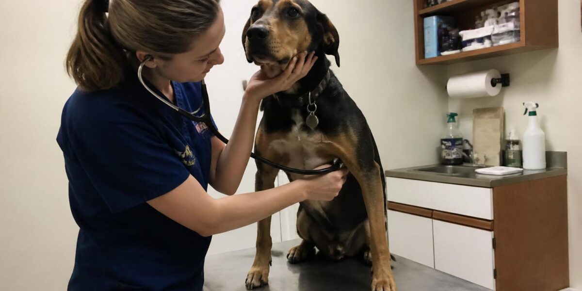
A mass in the front of the chest can stir real worry, and one of the tumors that develops there is the thymoma. What makes thymoma dogs cats develop especially interesting, and sometimes confusing, is that the trouble it causes often comes not from the tumor itself but from the strange body-wide effects it can trigger, known as paraneoplastic syndromes. A dog might first be seen for difficulty swallowing or weakness rather than for a chest mass. Understanding this tumor helps you connect seemingly unrelated signs and pursue the right diagnosis.
In this article
This guide explains what a thymoma is, the paraneoplastic problems it can cause, how it is distinguished from a common look-alike, and how it is treated.
Thymoma Dogs Cats and Owners Should Know About
The thymoma dogs cats and other companion animals develop is essentially the same disease across species, differing mainly in some of the paraneoplastic effects it tends to trigger. Understanding the basics of the tumor first makes those species-specific differences easier to follow.
The thymus is an organ in the cranial mediastinum, the space in the front of the chest between the lungs. It is important to the developing immune system, and in young animals it is sizable before gradually shrinking with age. A thymoma is a tumor arising from the epithelial cells of the thymus.
Thymomas are typically slow-growing and may be non-invasive or invasive. Non-invasive thymomas remain encapsulated and are more amenable to complete surgical removal, while invasive thymomas grow into surrounding structures, making treatment more challenging. They occur in both dogs and cats, generally in older animals.
As a thymoma enlarges within the confined space of the chest, it can begin to press on the lungs, the heart, and the major vessels, sometimes causing coughing, exercise intolerance, or labored breathing. Some animals also accumulate fluid in the chest. These local effects, combined with the paraneoplastic syndromes described below, mean that the same tumor can present in strikingly different ways from one patient to the next, which is part of what makes thymoma such an interesting and sometimes elusive diagnosis.
The Paraneoplastic Connection
Thymomas are notable for the paraneoplastic syndromes they can trigger, which are body-wide effects driven by the tumor rather than by direct local invasion. These effects are often what bring the animal to the veterinarian in the first place. Our overview of paraneoplastic syndromes in pets provides the broader context for why these patterns are so important to recognize.
The most clinically important thymoma-associated syndromes include:
- Myasthenia gravis — an immune-mediated disorder of nerve-to-muscle signaling that causes muscle weakness.
- Hypercalcemia — elevated blood calcium, which can affect the kidneys and overall health.
- Exfoliative dermatitis — a scaly, flaky skin condition seen particularly in cats.
Myasthenia Gravis and Its Dangers
Myasthenia gravis is the most serious paraneoplastic syndrome linked to thymoma because of its complications. The immune system produces antibodies that interfere with signaling at the junction between nerves and muscles, causing weakness that worsens with activity.
A particularly dangerous consequence is megaesophagus, an enlarged, poorly functioning esophagus that cannot move food effectively into the stomach. This leads to regurgitation and a high risk of aspiration pneumonia, a potentially life-threatening lung infection. In some dogs, the megaesophagus and its complications are more immediately threatening than the thymoma itself, which is why recognizing the link is so important.
Hypercalcemia and Other Effects
Some thymomas drive up blood calcium levels, producing hypercalcemia of malignancy. Signs include increased thirst and urination, lethargy, and reduced appetite, and severely elevated calcium can damage the kidneys. Our article on hypercalcemia of malignancy in dogs explains how this is recognized and managed, sometimes as an emergency requiring a board-certified critical care specialist (a Diplomate of the American College of Veterinary Emergency and Critical Care, or DACVECC).
In cats, thymoma is a recognized cause of exfoliative dermatitis, in which the skin becomes scaly, crusty, and flaky over much of the body. This unusual skin presentation can be the clue that prompts a search for an underlying chest mass.
Distinguishing Thymoma from Mediastinal Lymphoma
A critical diagnostic challenge is that thymoma is not the only tumor that forms a mass in the cranial mediastinum. Mediastinal lymphoma also occurs there, and the two are treated completely differently, so telling them apart is essential.
Diagnosis combines imaging (radiographs, ultrasound, or CT) with sampling. On cytology, a thymoma typically yields a mix of epithelial cells and many small mature lymphocytes, sometimes with mast cells, whereas lymphoma yields a uniform population of immature lymphocytes. A board-certified pathologist’s interpretation is key. Our articles on mediastinal lymphoma in cats and feline mediastinal lymphoma and chylothorax management describe that look-alike in detail, underscoring why an accurate diagnosis must come before treatment.
The reason this distinction is so consequential is that the two diseases call for opposite first steps. Lymphoma is treated with chemotherapy and generally does not start with surgery, whereas a non-invasive thymoma is best removed surgically. Operating on what turns out to be lymphoma, or starting chemotherapy for what is actually a resectable thymoma, would be a serious misstep. When cytology alone leaves doubt, additional tests such as flow cytometry or a tissue biopsy may be needed to settle the question before any treatment begins.
Treatment Options
Surgical removal is the treatment of choice for non-invasive thymomas. Complete resection of an encapsulated tumor can be curative and offers the best long-term outlook. This is major thoracic surgery best performed by a board-certified surgeon (a Diplomate of the American College of Veterinary Surgeons, or DACVS), often with careful anesthetic management given the chest location and any concurrent myasthenia gravis.
For invasive thymomas that cannot be completely removed, radiation therapy can help control the tumor and is overseen by a board-certified radiation oncologist (DACVR-RO). Any chemotherapy or drug decisions belong to a board-certified medical oncologist (a Diplomate of the American College of Veterinary Internal Medicine, oncology specialty, or DACVIM-Oncology) working with your primary veterinarian; we do not provide doses here. Managing associated myasthenia gravis and aspiration pneumonia is a parallel priority, sometimes more urgent than the tumor itself.
Prognosis and What to Expect
Outlook depends largely on whether the tumor is invasive and on the presence and severity of paraneoplastic syndromes. Among thymoma dogs cats and other patients, those with non-invasive tumors that are completely removed surgically can do very well, sometimes with long survival. Invasive tumors carry a more guarded prognosis, and the presence of megaesophagus with aspiration pneumonia worsens the outlook because of its complications.
Any survival figure should be understood as a median with wide individual variation and never as a prediction for a specific patient. The completeness of surgery, the control of paraneoplastic effects, and the animal’s overall health all matter. A frank conversation with your veterinary team about realistic goals will guide the best plan. For general grounding, our overview of cancer in dogs may be helpful.
Cost, Access, and Adoption Perspective
Advanced imaging, thoracic surgery, radiation, and management of complications such as aspiration pneumonia each add cost, and these vary by region, program, and specialist; figures reflect 2024 to 2025 ranges. Financing through CareCredit or Scratchpay, and grant programs such as The Pet Fund, may help. Surgeons, oncologists, and radiation oncologists with this expertise concentrate at referral centers, so your primary veterinarian may coordinate care remotely if a specialist is not nearby.
An animal treated successfully for a non-invasive thymoma can return to a full life, and such a history should not discourage a loving adoption. Many families care for pets with treated conditions and find the bond all the more rewarding for it. If you are considering a pet with a chronic or previously treated condition, our guide to adopting a pet with a chronic illness offers encouraging, practical advice.
Frequently Asked Questions
What is a thymoma?
A thymoma is a tumor arising from the thymus, an immune organ in the front of the chest. It occurs in both dogs and cats, usually older ones, and is often slow-growing. It can be non-invasive and encapsulated or invasive into surrounding tissue, which affects treatment and outlook.
Why might my pet show weakness or trouble swallowing?
Thymomas can trigger myasthenia gravis, an immune disorder that disrupts nerve-to-muscle signaling and causes weakness. It can also cause megaesophagus, leading to regurgitation and a risk of aspiration pneumonia. These paraneoplastic effects are sometimes more urgent than the tumor itself and are an important clue to the diagnosis.
How is thymoma told apart from lymphoma?
Both can form a mass in the front of the chest, but they are treated very differently. Cytology helps distinguish them, as thymoma shows a mix of epithelial cells and mature lymphocytes while lymphoma shows uniform immature lymphocytes. A board-certified pathologist’s interpretation is essential before treatment begins.
Can a thymoma be cured?
A non-invasive thymoma that is completely removed surgically can sometimes be cured, with good long-term outcomes. Invasive tumors are harder to remove fully and carry a more guarded prognosis. Your veterinary team can give a realistic assessment based on imaging and the tumor’s behavior.
What is the danger of aspiration pneumonia?
When megaesophagus develops alongside thymoma-associated myasthenia gravis, food and fluid can be inhaled into the lungs, causing aspiration pneumonia. This lung infection can be life-threatening and sometimes poses a more immediate risk than the tumor, so it requires prompt and careful management.


