
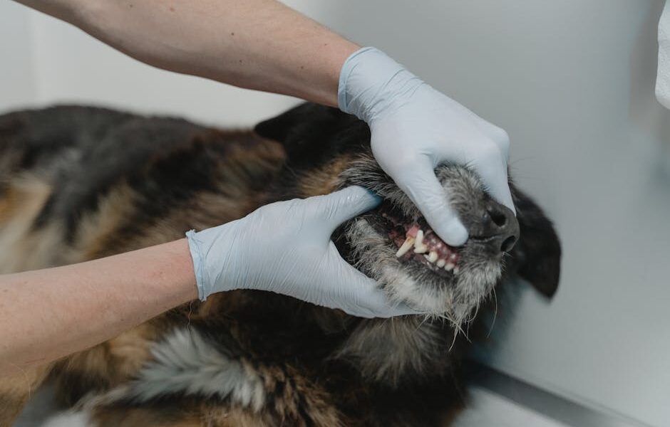
Yes, Dogs Can Get True Cavities
Canine dental caries — true cavities caused by bacterial demineralization of enamel and dentin — are a real diagnosis in dogs, even though they are notably less common than in humans. Surveys put the prevalence in pet dogs at roughly 5%, with some breed and diet groups higher. This is one of those topics where general internet wisdom (“dogs do not get cavities”) is wrong because the speaker is mixing up “less common” with “does not exist.” Your dog can get a cavity. It can require restoration or extraction. And it can be missed without dental radiographs.
In this article
- Yes, Dogs Can Get True Cavities
- The Bacteriology: Streptococcus mutans and Friends
- Which Teeth Get Cavities (and Why)
- How a Carious Lesion Is Diagnosed
- Restoration vs Extraction: The Decision
- Anesthesia and Diagnostic Workup Matter
- Diet’s Role in Canine Caries Risk
- Prevention: Honest, Achievable Targets
- What to Watch for at Home
- Distinguishing Caries from Other Tooth Discolorations
- Frequently Asked Questions
Two factors protect dogs from the runaway cavity problem we see in human dentistry: their saliva is more alkaline (pH around 7.5 vs human 6.5), which neutralizes acid-producing bacteria, and their teeth have wider interdental spaces and more conical crowns that resist plaque retention in the pit-and-fissure pattern. But these protections are partial, not absolute. When the diet shifts toward fermentable carbohydrates and a dog has the wrong tooth anatomy in the wrong place, you get caries.
This guide covers the bacteriology, the at-risk teeth, the diagnostic workup, and the decision tree between restoration and extraction. For the broader dental care framework, see our pieces on periodontal disease staging and the AVDC home brushing protocol.
The Bacteriology: Streptococcus mutans and Friends
The classic caries pathogen in humans is Streptococcus mutans, which ferments dietary carbohydrates into lactic acid and demineralizes enamel below pH 5.5. The canine oral microbiome differs from the human one — dogs carry more gram-negative anaerobes and fewer S. mutans species — but S. mutans, S. salivarius, and Actinomyces species have all been recovered from canine carious lesions. The mechanism is the same: bacterial biofilm + fermentable carbohydrate substrate + susceptible tooth surface = demineralization.
What is different in dogs is the carbohydrate exposure. Wild and ancestral dog diets contain almost no fermentable sugar; modern commercial dry kibble carries variable starch content, and dogs eating table scraps, dental treats with hidden sweeteners, or human dessert foods get repeated acidic exposure that their oral defenses were not evolved for. (Never share xylitol-containing human treats — see xylitol toxicity in dogs for why even a small amount can be lethal.)
Which Teeth Get Cavities (and Why)
Caries in dogs almost always strike the maxillary first molars (the carnassial teeth on the upper jaw) and occasionally the lower first molars. These teeth have a flatter occlusal surface with deep pit-and-fissure anatomy where plaque and food debris can sit for hours, exactly the pattern that traps fermentable carbohydrate and bacterial biofilm.
Less commonly, smooth-surface caries develop on the buccal (cheek) surfaces of premolars, particularly in dogs with mouth conformations that allow food impaction between teeth. Smooth-surface caries are easier to brush away in early stages, which is why the home brushing protocol genuinely matters for prevention.
Dogs at higher risk include:
- Breeds with crowded or rotated teeth (brachycephalics like Boxers, Pugs, Shih Tzus)
- Dogs fed soft, sticky foods or frequent carbohydrate-rich treats
- Dogs with chronic xerostomia (dry mouth) from medication or salivary gland disease
- Dogs with prior dental restorations that have failed at the margin
How a Carious Lesion Is Diagnosed
Visual examination under anesthesia is the starting point: the dental clinician probes the suspicious tooth with a sharp explorer, looking for a sticky catch in the pit or fissure. A positive “stick” suggests demineralized enamel that has not yet cavitated through. Dental radiographs then confirm the depth — has the lesion reached dentin, is it close to pulp, or has it already perforated into the pulp chamber?
Five stages of caries (G.V. Black classification, adapted for dogs):
- Stage 1: Demineralization confined to enamel
- Stage 2: Lesion into dentin, no pulp involvement
- Stage 3: Lesion close to pulp but not exposed
- Stage 4: Pulp exposure, vital tooth
- Stage 5: Pulp necrosis, non-vital tooth, possible periapical involvement
This is why the dental x-rays for dogs standard matters — a clinician staging caries without radiographs is guessing about pulp distance, and guessing wrong means either over-treating (unnecessary root canal) or under-treating (filling that fails when pulpitis develops a year later).
Restoration vs Extraction: The Decision
For Stage 1-2 lesions where the pulp is not exposed and adequate tooth structure remains, restoration with a glass ionomer or composite resin filling is feasible. The dental team isolates the tooth, removes the carious dentin with a slow-speed bur, etches and bonds the cavity walls, places the restoration, and finishes the margin. Done well, restorations last 5-10 years.
For Stage 3-4 lesions, the decision is harder. If the pulp is exposed but the tooth is vital and the patient is a good candidate, a vital pulp therapy (direct pulp cap) or full root canal followed by restoration may save the tooth. These procedures require a DAVDC board-certified veterinary dentist and have failure rates that owners must understand before consenting.
For Stage 5 (non-vital tooth with apical disease) or any patient where ongoing dental care will not be maintained, extraction is the more honest choice. A failed restoration in 18 months is worse than a clean extraction now, because the tooth has been hurting that whole time. Talk to your dental team about realistic home care commitment before choosing restoration over extraction.
Anesthesia and Diagnostic Workup Matter
Any restoration, vital pulp therapy, or root canal in dogs requires general anesthesia — there is no awake, cooperative, or “anesthesia-free” route to a properly bonded composite filling. Dogs do not hold still for rubber dam isolation and acid etching. Read why anesthetic-free dental claims are not credible and why the modern anesthesia protocol makes cleaning under anesthesia genuinely low-risk.
Pre-anesthesia bloodwork, ASA status assessment, and intraoperative monitoring (BP, ECG, capnography, pulse-ox, temperature) are now standard of care. Senior dogs benefit from the senior pet pre-anesthesia workup to identify cardiac, renal, or hepatic concerns before the procedure.
Diet’s Role in Canine Caries Risk
The relationship between diet and canine dental caries deserves more attention than it usually gets. The fermentable-carbohydrate hypothesis — bacterial fermentation of dietary sugars producing acid that demineralizes enamel — is well-established in human dentistry and increasingly recognized in veterinary dentistry. Dogs eating diets high in soft, sticky, sugar-containing treats (frosted dog biscuits, semi-moist treats, table scraps including bread and fruit) get more cumulative acidic challenge than dogs on lower-carbohydrate diets.
This does not mean grain-free or raw diets are caries-protective. The fermentable substrate matters, not the gluten content. Dogs on raw or fresh-cooked diets that include rice, sweet potato, or fruit can still get caries. The cleanest practical advice: minimize sticky, sweet treats; keep dental treats to VOHC-accepted products that have been tested for plaque effects rather than marketed on appearance; and avoid sharing human dessert foods.
Prevention: Honest, Achievable Targets
Two interventions move the needle on caries prevention:
- Daily home brushing with enzymatic dog toothpaste using the AVDC-accepted technique. Mechanical plaque removal is the single most effective intervention.
- Annual professional cleaning under anesthesia with full-mouth radiographs. This catches early lesions when restoration is still possible.
VOHC-accepted dental chews can be useful adjuncts, but no chew has been demonstrated to prevent caries the way mechanical brushing does. Diet matters: reduce sticky, sugary treats and human dessert sharing. Avoid bones, ice cubes, and tug-of-war on hard toys that can chip enamel and create new colonization sites. For broader brushing technique guidance, see also how to brush dog teeth.
What to Watch for at Home
Dogs hide oral pain. Signs that something is wrong with a tooth include dropping food, chewing on one side, sudden disinterest in hard treats, pawing at the mouth, halitosis (bad breath that is worse than the dog’s baseline), and visible discoloration of a tooth crown (dark spot or pink crown). Any of these warrants a vet exam with anesthetized dental radiographs — not a hopeful retail “dental treat” purchase.
The behavioral changes that signal oral pain often arrive before the visible ones. Dogs with developing caries may become slightly more irritable during play, less enthusiastic about meals they previously loved, more selective about toy choices (avoiding hard rubber for soft plush), or briefly stop and reposition during chewing. These small shifts are easy to attribute to “getting older” or “just not in the mood” when they are actually pain reports. Take them seriously.
For broader oral disease that may coexist with caries, our reference articles on gingivitis in dogs and tooth root abscess are good companions. Caries lesions and periodontal disease often coexist, particularly in dogs with crowded dentition.
Distinguishing Caries from Other Tooth Discolorations
Not every dark spot on a dog’s tooth is a cavity. Other causes of crown discoloration that owners and even some clinicians confuse with caries include:
- Extrinsic staining from food, water, and chewing habits — surface-only, removed by professional polishing
- Intrinsic discoloration from trauma — pulpal hemorrhage shows as a pink, gray, or purple crown reflecting blood breakdown products in the dentinal tubules
- Enamel hypoplasia from puppyhood febrile illness (canine distemper historically a classic cause) — pitted enamel that may look like a caries lesion but represents developmental defect
- Tetracycline staining from antibiotic exposure during tooth development — yellow-brown bands across multiple teeth
The dental exam under anesthesia distinguishes these by visual inspection plus explorer probe — a true caries lesion catches the explorer in the demineralized pit; staining does not. Dental radiographs further distinguish by showing dentin lucency in caries but not in pure staining.
Frequently Asked Questions
How common are cavities in dogs really?
Studies put true caries prevalence at around 5% of adult pet dogs, but anyone using the visual-only standard will under-diagnose. With dental radiographs, the rate is likely higher. The disease is real and missed often.
Will dental chews prevent cavities?
VOHC-accepted chews are tested against plaque and calculus, not caries specifically. They likely reduce caries risk by reducing plaque burden, but no chew is a substitute for daily brushing. The VOHC evidence review covers full nuance.
Can my dog eat sugary treats?
Avoid sticky carbohydrate-rich human treats. Avoid xylitol-containing products entirely. Small amounts of plain, unsweetened fruit and vegetable treats are fine for systemic health, but anything that sticks to teeth raises caries risk.
Does a restoration last forever?
No. Composite restorations in dogs typically last 5-10 years. Restoration margins can fail, recurrent caries can develop, and the underlying tooth can fracture. Plan on professional dental evaluation every 6-12 months after any restoration.
Should I see a board-certified veterinary dentist?
For root canals, vital pulp therapy, and any restoration in a working or service dog where preserving the tooth has high stakes, yes. For simple extractions of carious teeth, your general-practice veterinarian is usually fully capable.


