
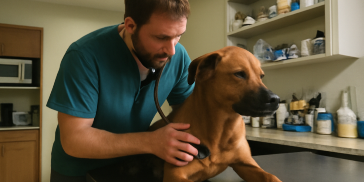
What Protein-Losing Enteropathy Means
Protein-losing enteropathy (PLE) in dogs is a serious condition where the gastrointestinal tract leaks blood proteins, especially albumin, into the gut. Normally, the intestinal wall keeps proteins safely in the bloodstream while absorbing nutrients from food. In PLE, that barrier breaks down, and proteins escape faster than the body can replace them.
In this article
- What Protein-Losing Enteropathy Means
- Recognizing the Signs
- The Diagnostic Workup
- Intestinal Lymphangiectasia Explained
- Diet as the Cornerstone of Treatment
- Medication Options
- Managing Fluid Accumulation
- Monitoring Long-Term
- Concurrent Conditions and Complications
- Supportive Care at Home
- Long-Term Outlook
- Frequently Asked Questions
Protein losing enteropathy dogs face is not a single disease but a syndrome that can result from several underlying causes. The most common include inflammatory bowel disease, intestinal lymphangiectasia (a condition affecting lymph drainage in the gut), intestinal lymphoma, severe parasitic infections, and other infiltrative diseases. Identifying the underlying cause guides treatment and prognosis.
The condition tends to be more serious than uncomplicated IBD because the protein loss has cascading effects throughout the body. Low albumin reduces blood pressure-holding capacity, allowing fluid to leak into the abdomen, chest, or under the skin. This combination makes PLE one of the more challenging GI conditions to manage.
Recognizing the Signs
Early signs often look similar to standard chronic GI disease: intermittent diarrhea, weight loss, decreased appetite, and sometimes vomiting. As protein loss worsens, more dramatic findings appear. Fluid accumulation in the abdomen (ascites) creates a distended belly. Chest fluid (pleural effusion) causes labored breathing. Limb swelling (edema) may appear in the legs.
Other signs include lethargy, weakness, exercise intolerance, and a generally unwell appearance. Some dogs lose significant muscle mass, especially over the spine and shoulders. The coat may become dull, and minor cuts or scrapes may bleed more than expected because clotting factors are also lost.
Several breeds are reported as predisposed to specific forms of PLE, including Yorkshire Terriers, Soft Coated Wheaten Terriers, Norwegian Lundehunds, and Basenjis. Mixed-breed dogs can develop PLE too, especially when underlying GI disease progresses. Any dog with chronic GI signs and weight loss should be evaluated for possible PLE.
The Diagnostic Workup
Diagnosis combines bloodwork, urinalysis, fecal testing, imaging, and ultimately biopsy. Bloodwork typically shows low albumin and often low globulin (panhypoproteinemia) along with low cholesterol, low calcium, and sometimes lymphopenia. These findings together strongly suggest PLE.
Other causes of low albumin must be ruled out, including liver disease (which decreases albumin production) and kidney disease (which loses albumin in urine). A urine protein-to-creatinine ratio rules out renal protein loss. Liver function tests assess hepatic albumin production. If these are normal, GI loss is the likely source.
Imaging with abdominal ultrasound assesses bowel wall thickness, lymph node enlargement, and any masses or fluid accumulation. Biopsy via endoscopy or surgery is usually needed to identify the underlying cause: IBD, lymphangiectasia, lymphoma, or other infiltrative disease. The biopsy guides specific treatment.
Intestinal Lymphangiectasia Explained
Lymphangiectasia is a condition where the lymphatic vessels in the intestinal wall become dilated and leaky. Lymph fluid, which is rich in proteins and fat, escapes into the gut lumen. The result is significant protein and fat loss along with the typical PLE picture.
The condition can be congenital (present from birth) or acquired due to chronic inflammation that obstructs lymph flow. Yorkshire Terriers and Soft Coated Wheaten Terriers are particularly predisposed. Treatment focuses on reducing fat in the diet (so less lymph is produced) and addressing any underlying inflammation.
Lymphangiectasia often responds particularly well to ultra-low-fat diets, sometimes combined with corticosteroids for any concurrent inflammation. The diet must be very strict because even small amounts of fat can trigger lymph leakage. Many dogs with lymphangiectasia stabilize beautifully on appropriate dietary management.
Diet as the Cornerstone of Treatment
Diet is the foundation of PLE management regardless of the underlying cause. Most dogs benefit from a highly digestible, low-fat diet that minimizes irritation to the gut and reduces lymph production. Therapeutic diets formulated for GI disease are usually the starting point, with ultra-low-fat versions for lymphangiectasia.
Hydrolyzed protein diets or novel protein diets may also be tried, especially when food allergy or IBD is suspected as the underlying cause. Some dogs respond best to home-prepared diets formulated by a veterinary nutritionist. Trial and adjustment may be needed to find the best fit.
Treats and table scraps are off the menu. Even small amounts of fatty or unfamiliar foods can disrupt the careful balance. Educate everyone in the household about what your dog can eat, and use prescription kibble pieces as treats when possible. Consistency makes the difference.
Medication Options
Corticosteroids like prednisone are commonly used to reduce gut inflammation, especially when IBD is the underlying cause. The dose is gradually tapered as symptoms improve and albumin levels rise. Some dogs need long-term low-dose steroid therapy to maintain remission.
Other immunosuppressants are added when steroids alone are insufficient or cause too many side effects. Cyclosporine, azathioprine, chlorambucil, and mycophenolate are all options. The choice depends on the underlying disease and individual response. Your veterinarian or a board-certified internist will guide selection.
Supportive medications include B12 (cobalamin) supplementation (almost always needed in PLE), antibiotics in some cases, and anti-clotting medications when blood proteins involved in clotting are very low. Plasma transfusions may be needed in severe cases to temporarily boost albumin. Monitoring guides each adjustment.
Managing Fluid Accumulation
Severe PLE often causes fluid accumulation in the abdomen, chest, or limbs because low albumin cannot hold fluid in the blood vessels. Mild ascites may not need direct treatment, but significant fluid accumulation interferes with breathing and comfort.
Diuretics can help in some cases, but they must be used cautiously because they can cause dehydration and electrolyte imbalances. Therapeutic abdominocentesis (draining fluid from the abdomen) provides quick relief but is a temporary fix. The long-term solution is raising albumin levels by treating the underlying cause.
Plasma transfusions provide donor albumin and clotting factors, which can stabilize critically ill dogs while definitive treatment takes effect. Synthetic colloids like hetastarch are used in some cases. These interventions require hospitalization and careful monitoring.
Monitoring Long-Term
Regular monitoring is essential. Your veterinarian will track weight, body condition, appetite, GI symptoms, and bloodwork (especially albumin, globulin, cholesterol, and calcium). Frequent visits early in treatment shift to less frequent monitoring once disease is stable.
At-home monitoring helps too. Track stool consistency, appetite, energy, weight, and any signs of fluid accumulation. Photographs of the belly can help identify subtle changes in distension. A simple weekly log gives your vet useful trend data.
Trends matter more than single numbers. Slowly rising albumin, stable weight, and good appetite are encouraging. Falling albumin or new fluid accumulation triggers a closer look and possible treatment adjustment. Periodic ultrasound rechecks may be recommended.
Concurrent Conditions and Complications
Dogs with PLE are at increased risk for blood clots because the proteins lost include those involved in blood clot regulation. Pulmonary thromboembolism is a serious complication that can cause sudden breathing difficulty. Some dogs are placed on anti-clotting medications as a precaution.
Calcium and vitamin D deficiencies can develop because both are bound to albumin and lost in PLE. Supplementation may be needed in severe cases. Bone changes are uncommon but possible in chronic disease, so attention to calcium balance matters.
Concurrent IBD or lymphoma must be managed alongside PLE. Our guide to IBD in dogs and lymphoma in dogs guide cover these companion conditions in depth.
Supportive Care at Home
Day-to-day care for a dog with PLE focuses on consistency and observation. Stick to the prescribed diet without exception, including treats and any flavored medications. Family members, dog walkers, and visitors all need to understand the rules so well-meaning extras do not derail the plan.
Watch for signs of fluid accumulation by gently feeling the belly each day. A subtly distended or “doughy” abdomen can be an early signal of returning ascites. Limb swelling, especially around the lower legs and feet, is another early sign. Photos and weekly weighings help you track changes over time.
Activity should be gentle and consistent rather than vigorous. Short leash walks and calm play maintain muscle tone without overtaxing a body that is short on protein and clotting factors. Avoid rough play with other dogs during fragile periods. Stress reduction supports healing and reduces flare risk.
Long-Term Outlook
Prognosis depends heavily on the underlying cause and response to treatment. Lymphangiectasia and IBD-related PLE often respond well to dietary and medical management, with many dogs achieving stable disease for years. Lymphoma-related PLE has a more guarded prognosis but can also respond to chemotherapy.
The earlier PLE is diagnosed and treated, the better the outcome. Severe cases with significant ascites, very low albumin, or complications carry a more guarded prognosis. Open communication with your veterinary team about goals and quality of life guides decisions over time.
For more on related canine wellness topics, our dog care blog has resources. If you are considering adopting a dog with chronic GI issues, the dog adoption hub offers guidance on welcoming a pet with manageable medical needs.
Frequently Asked Questions
Is PLE in dogs curable?
PLE is generally not curable but is often manageable. Many dogs achieve good control of underlying causes with diet, medications, or both, and live comfortable lives. Some have flares requiring adjustments. Lifelong monitoring and a thoughtful treatment plan are essential.
How long until my dog’s albumin levels improve?
Improvement may begin within weeks of starting appropriate treatment, but full normalization can take months. The pace depends on the underlying cause and how strictly the diet and medications are followed. Periodic bloodwork tracks progress.
Can my dog still have any treats with PLE?
Yes, but treats must match the prescribed diet. Most therapeutic GI diet brands offer matching treats, and ultra-low-fat options exist for dogs with lymphangiectasia. Forbidden treats can sabotage results, so always check with your veterinarian before introducing new options.
Are some breeds predisposed to PLE?
Yes. Yorkshire Terriers, Soft Coated Wheaten Terriers, Norwegian Lundehunds, and Basenjis are reported as predisposed. Mixed-breed dogs can develop PLE too, especially when underlying GI disease progresses without treatment. Awareness in predisposed breeds helps with early diagnosis.
When should I take my dog to the vet?
Call promptly for chronic diarrhea, weight loss, decreased appetite, or any swelling of the belly or limbs. Severe lethargy, breathing difficulty, or signs of clotting problems are urgent. Always consult your veterinarian for diagnosis and ongoing PLE management.


